Furthermore, the Journal is also present in Twitter and Facebook. Manuscripts will be submitted electronically using the following web site:, link which is also accessible through the main web page of Archivos de Bronconeumologia.Īccess to any published article, is possible through the Journal's web page as well as from PubMed, Science Direct, and other international databases. The Journal is published monthly in English. It is a monthly Journal that publishes a total of 12 issues and a few supplements, which contain articles belonging to the different sections.Īll the manuscripts received in the Journal are evaluated by the Editors and sent to expert peer-review while handled by the Editor and/or an Associate Editor from the team. Other types of articles such as reviews, editorials, a few special articles of interest to the society and the editorial board, scientific letters, letters to the Editor, and clinical images are also published in the Journal. Una MRI con medio de contraste es la mejor manera de ver los tumores cerebrales.Archivos de Bronconeumologia is a scientific journal that preferentially publishes prospective original research articles whose content is based upon results dealing with several aspects of respiratory diseases such as epidemiology, pathophysiology, clinics, surgery, and basic investigation. La MRI es muy buena para detectar y localizar algunos tipos de cáncer. La MRI crea imágenes de partes del tejido blando del cuerpo que a veces son difíciles de ver usando otros estudios por imágenes. Una MRI puede tomar cortes transversales (vistas) desde muchos ángulos, como si alguien estuviera mirando una sección de su cuerpo de frente, de costado, o por encima de su cabeza. Sin embargo, la MRI usa imanes muy potentes en lugar de radiación para crear las imágenes. These methods share the need for acquisition methods with high temporal and spatial resolution.Īl igual que la CT, la imagen por resonancia magnética (MRI) crea imágenes transversales de sus órganos internos. Quantitative DCE-MRI requires special data acquisition techniques and analysis software.Ĭontrast enhanced magnetic resonance angiography (CE-MRA) allows the visualization of vessels and the temporal resolution provides a separation of arteries and veins. DCE-MR imaging is used for diagnosis of cancer (see also liver imaging, abdominal imaging, breast MRI, dynamic scanning) as well as for diagnosis of cardiac infarction (see perfusion imaging, cardiac MRI). The focus of dynamic contrast enhanced MRI (DCE-MRI) is on contrast kinetics with demands for spatial resolution dependent on the application.

Some methods are available to improve the contrast of different tissues.

The need to more accurately characterize different types of lesions and to detect all malignant lesions is the main reason for the use of intravenous contrast agents.

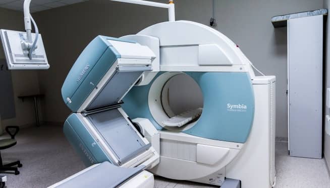
Spanish translation: Resonancia magnética con contrasteĬontrast enhanced MRI is a commonly used procedure in magnetic resonance imaging.


 0 kommentar(er)
